
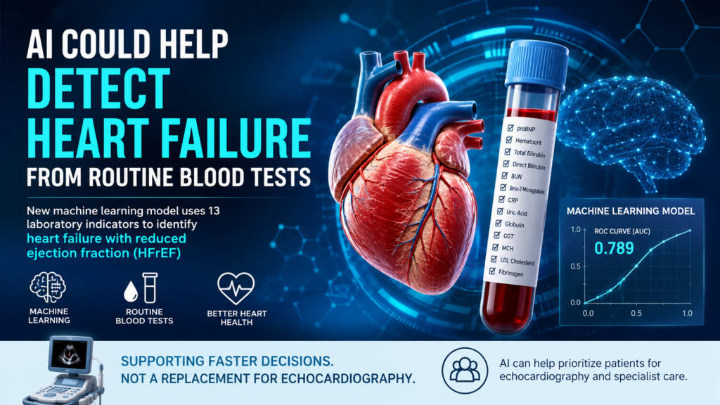
A new machine learning model could help doctors identify heart failure with reduced ejection fraction (HFrEF) using information from routine laboratory blood tests, potentially helping prioritize patients for echocardiography when cardiac imaging is not immediately available.
The study, published in Frontiers in Cardiovascular Medicine, evaluated whether commonly available laboratory indicators could be combined with machine learning to distinguish HFrEF from other forms of heart failure, including heart failure with mildly reduced ejection fraction (HFmrEF) and heart failure with preserved ejection fraction (HFpEF).
The findings highlight a growing role for artificial intelligence and machine learning in heart failure diagnosis and risk assessment, although researchers emphasize that the model is not intended to replace echocardiography or clinical evaluation.
What Is Heart Failure With Reduced Ejection Fraction?
Heart failure occurs when the heart cannot pump or fill with blood effectively enough to meet the body’s needs.
One important way doctors classify heart failure is by measuring the left ventricular ejection fraction (LVEF). HFrEF generally refers to heart failure in which the left ventricular ejection fraction is 40% or less. HFmrEF is generally classified as an ejection fraction of 41% to 49%, while HFpEF refers to an ejection fraction of 50% or higher.
Ejection fraction is commonly evaluated using an echocardiogram, which provides information about how effectively the heart is pumping.
The American Heart Association notes that a typical ejection fraction is approximately 55% to 70%, although ejection fraction is only one part of evaluating heart function.
How Could Machine Learning Help Detect HFrEF?
The new research explored whether routine blood-test results could provide enough information for a machine learning system to identify patients more likely to have HFrEF.
Researchers from Guangxi Medical University and Guigang City People’s Hospital analyzed medical records from 1,480 hospitalized patients with chronic heart failure.
Among them:
- 377 patients had HFrEF
- 1,103 patients had HFmrEF or HFpEF
- Laboratory measurements were collected from the first blood test within 24 hours of admission
- Echocardiography was generally performed within 48 hours
- Researchers evaluated six different machine learning algorithms
The study used a 7:3 training-to-testing split and applied a machine learning pipeline involving preprocessing, feature selection, model training and evaluation.
The Model Used 13 Routine Laboratory Indicators
After feature selection, researchers identified 13 laboratory indicators for the final model.
These included:
- proBNP
- Hematocrit (HCT)
- Total bilirubin
- Direct bilirubin
- Blood urea nitrogen (BUN)
- Beta-2 microglobulin
- C-reactive protein (CRP)
- Uric acid
- Globulin
- Gamma-glutamyl transferase (GGT)
- Mean corpuscular hemoglobin (MCH)
- LDL cholesterol
- Fibrinogen
The researchers said the indicators represented several biological processes, including cardiac stress, renal function, inflammation, hematologic status, hepatobiliary congestion, lipid metabolism and coagulation.
Among these variables, proBNP emerged as the dominant predictor in the study’s SHAP analysis, followed by hematocrit, direct bilirubin, globulin, uric acid, GGT, total bilirubin and LDL cholesterol.
Random Forest and XGBoost Delivered the Best Results
Researchers tested six machine learning approaches, including:
- Random forest
- XGBoost
- Logistic regression
- Support vector machine
- K-nearest neighbors
- Multilayer perceptron
In the independent test set, random forest and XGBoost both achieved an area under the ROC curve (AUC) of 0.789.
Logistic regression produced a similar AUC of 0.784.
The random forest model also had the lowest Brier score, a measure related to prediction accuracy and calibration, at 0.152. However, statistical comparisons did not show a significant difference in AUC between the different models.
This is important because the results do not demonstrate that artificial intelligence has definitively outperformed conventional statistical approaches.
Instead, they suggest that machine learning can potentially combine multiple routine laboratory measurements into a useful risk-stratification tool.
Changing the Threshold Improved Sensitivity
One of the more interesting findings came from changing the decision threshold of the random forest model.
At its default threshold, sensitivity was relatively limited. Researchers therefore tested a lower threshold of 0.15.
At that threshold, sensitivity increased to 0.912, while the negative predictive value reached 0.935.
The researchers suggest that this type of threshold could potentially be useful for a rule-out triage strategy, helping determine which patients should receive prioritized echocardiography or specialist assessment.
However, a higher sensitivity can also result in more false-positive results. Therefore, the model should not be interpreted as providing a definitive diagnosis.
Why Echocardiography Still Matters
Despite the promising findings, machine learning cannot currently replace an echocardiogram for determining a patient’s ejection fraction.
Echocardiography remains an important method for assessing left ventricular function and classifying heart failure according to ejection fraction.
The researchers specifically describe their model as an adjunctive triage tool, rather than a substitute for echocardiographic phenotyping.
This distinction is particularly important for patients because heart failure treatment decisions can depend on the patient’s heart failure phenotype and overall clinical condition.
Potential Benefits of AI-Assisted Heart Failure Diagnosis
If validated in larger and more diverse patient populations, a laboratory-based machine learning model could have several potential advantages.
1. Faster patient prioritization
Blood tests are routinely performed in many hospitals. A predictive model could potentially identify patients who may benefit from faster cardiac imaging.
2. More efficient use of echocardiography
Where access to echocardiography is limited, an AI-assisted triage system could potentially help healthcare teams prioritize patients for imaging.
3. Use of existing medical information
The model relies on routine laboratory indicators rather than requiring an entirely new diagnostic test.
4. Support for clinical decision-making
Machine learning could potentially provide an additional source of information alongside symptoms, physical examination, laboratory testing and imaging.
These potential applications remain areas for future research rather than established clinical practice.
What Are the Limitations of the Study?
The findings should be interpreted cautiously.
The study was retrospective and conducted at a single hospital, using data from patients hospitalized between January 2019 and December 2022.
The researchers also acknowledged limitations related to the retrospective nature of the dataset, including heterogeneity in how ejection fraction measurements were obtained and the lack of complete information needed to separately identify patients with heart failure with improved ejection fraction.
Most importantly, the model has not yet undergone the kind of broad external and prospective validation needed before it could be considered reliable across different hospitals, populations and healthcare systems.
The researchers therefore state that the model should be considered an adjunctive triage tool while further validation is performed.
AI Is Becoming a Major Research Area in Heart Failure
This study is part of a much broader movement toward artificial intelligence in cardiovascular medicine.
Recent research shows rapidly increasing interest in machine learning for heart failure diagnosis, prognosis, mortality prediction, phenotyping and treatment-response assessment. A recent bibliometric analysis found that “heart failure,” “machine learning” and “artificial intelligence” are among the most frequently occurring keywords in research on AI applications in heart failure.
Other 2026 research has also investigated AI for detecting left ventricular dysfunction and heart failure from electrocardiograms, demonstrating that machine learning is being explored across different types of cardiovascular data.
The broader direction is toward combining information from laboratory tests, ECGs, imaging and clinical records to improve cardiovascular risk assessment.
What Happens Next?
The next step for this type of technology is external validation.
Researchers will need to determine whether the model works reliably in:
- Different hospitals
- Different countries
- Larger patient populations
- Different ethnic and demographic groups
- Patients with different comorbidities
- Real-world prospective clinical settings
If future studies confirm the findings, machine learning could eventually become a useful support tool for identifying patients who need more urgent cardiac evaluation.
For now, however, the research represents an encouraging proof of concept rather than a replacement for established heart failure diagnostic methods.
The Bottom Line
A new machine learning model using 13 routine laboratory indicators showed moderate ability to distinguish heart failure with reduced ejection fraction from heart failure with mildly reduced or preserved ejection fraction.
Random forest and XGBoost achieved the highest test-set AUC of 0.789, while adjusting the random forest threshold increased sensitivity to 91.2% and negative predictive value to 93.5%.
The findings suggest that artificial intelligence could potentially help healthcare professionals prioritize patients for echocardiography, particularly when imaging is delayed or resources are limited.
But the model still requires external and prospective validation. An AI prediction should not be considered a diagnosis, and echocardiography remains essential for determining ejection fraction and heart failure phenotype.
Source and Study Details
The research was published in Frontiers in Cardiovascular Medicine on August 10, 2026.
Study: “Machine learning-based identification of heart failure with reduced ejection fraction using routine laboratory indicators”
Authors: Zhiping Meng, Xuezhan Qin, Binbin Liang, Wenpei Lin, Wentan Xie and Guinian Du.
DOI: 10.3389/fcvm.2026.1856570.
This article is for informational purposes and does not provide medical advice. Diagnosis and treatment decisions should be made by qualified healthcare professionals.